Multifunctional Imager YR06067–YR06068
Multifunctional Imager YR06067–YR06068 — Kalstein laboratory equipment with technical specifications, advanced features, and certified professional solutions for scientific use.
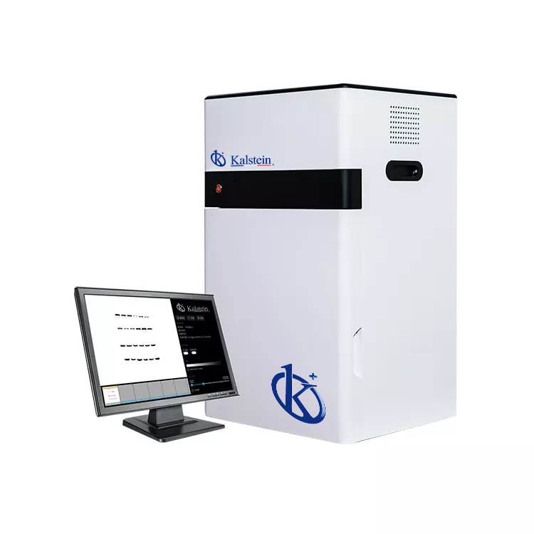
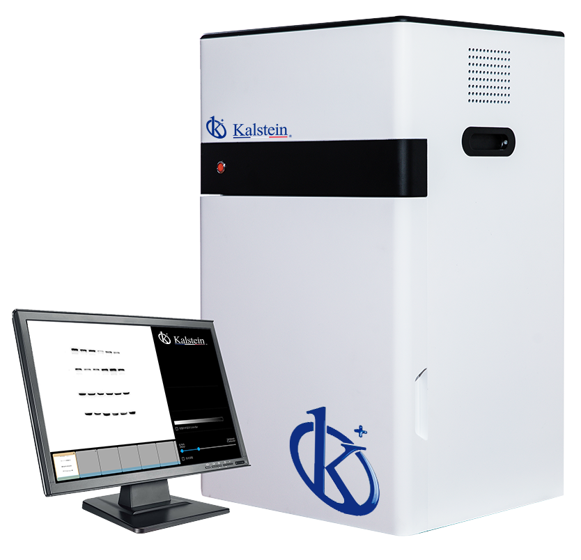
The Multifunctional Imager YR06067–YR06068 stands out with its impressive 20x20 cm view area and user-friendly design, making it an essential tool in laboratories. Its distinctive features, such as the standard Western blot imager with ECL and a cooled digital camera using a sensitive CCD chip, make it highly versatile. The imager’s compatibility with a wide range of optional accessories and its offer of unlimited, free-to-download, and regularly updated software maximize functionality, making it a reliable choice for various imaging needs.
Market Price
The average market price for advanced imaging systems such as the Multifunctional Imager YR06067–YR06068 typically ranges from $20,000 to $21,000. This reflects the state-of-the-art technology and robust features offered by this model. For accurate and up-to-date pricing, it is recommended to request a personalized quote. With Kalstein Plus, you can easily generate an automatic quote now!
Frequently Asked Questions
What is the view area of the imager? The Multifunctional Imager provides a large view area of 20x20 cm, perfect for various applications.
Is the software included? Yes, the imager comes with software that can be downloaded for free, providing unlimited use and free updates.
What camera technology does it use? This model uses a cooled CCD camera, vital for high-quality imaging results.
Advantages and Disadvantages
The Multifunctional Imager YR06067–YR06068 offers significant advantages such as superior image quality due to the sensitive CCD camera and extensive application range with its optional accessories. However, a potential downside is the initial investment cost, which might be high for some budgets, although its long-term benefits can outweigh this concern.
Product Use in the Field
In practical settings, the Multifunctional Imager is indispensable for imaging applications that require precision, such as Western blot analyses. The device's ability to handle large samples with clarity due to its cooling technology and extensive view area results in effective and efficient workflows in both research and clinical labs.
Recommendations
To get the best out of your Multifunctional Imager, ensure regular software updates and perform routine maintenance checks on the camera and lens systems. Consider training sessions for lab personnel to boost efficiency and reduce the learning curve associated with high-tech equipment.
Features
- Large view area (maximum 20x20cm)
- Easy to use
- Standard Western blot imager with ECL
- Wide application with optional accessories
- Cooled digital camera adopting sensitive CCD chip
- Unlimited Software, free download, free update.
Technical Specifications
| Model | YR06067 | YR06068 |
| Digital Camera | Cooled CCD Camera | |
| Physical Resolution | 6 Megapixels,2750*2200 | |
| Pixel Density and A/D | 16 Bit | |
| Grey Scales | 65536 | |
| Dynamic Range | ≥4.6OD | |
| Cooling Temperature | -60℃ below ambient, -30℃ regulated | |
| Scientific Lens (Difference between both models) | Sensitive F0.95 Lens | Ultra sensitive F0.8 Lens |
| Chemiluminescence sample tray | 4 Layer | |
| UV Transmission | 302nm-312nm, area:21x26cm | |
| White LED Transmission | Area: 19x26cm | |
| Filter Wheel | FIve position | |
| Filter | Gel Doc Filter | |
| UV Protection | UV Protection Shield | |
| Image Acquisition Software | No software dongle required, unlimited users, free update | |
| Image Analysis Software | No software dongle required, unlimited users, free update | |
Technical specifications
| Dimensions | L 50 × W 55 × H 88 cm |
|---|---|
| Weight | 29 kg |
| Manufacturer | Kalstein |
Available models
Frequently asked questions
How to know the prices of Multifunctional Imager YR06067–YR06068?
To know the price of Multifunctional Imager YR06067–YR06068, please send us an email with your request through the contact form AQUI.
What are the delivery times for Multifunctional Imager YR06067–YR06068?
Delivery time depends on stock availability and freight type (air or sea). In stock: air 15-30 days, sea 45-60 days. Out of stock: air 30-60 days, sea 60-90 days.
How to make a purchase of Multifunctional Imager YR06067–YR06068?
You can buy by email ([email protected]), phone (+33 (0) 1 70 39 26 50) or through the official Kalstein website in your country.
How does the warranty work for Multifunctional Imager YR06067–YR06068?
All Kalstein equipment comes with a 1-year warranty against manufacturing defects. The warranty does not cover damage from improper installation or misuse. See our «terms and conditions» AQUI.
Can I request a quote online for Multifunctional Imager YR06067–YR06068?
Yes, you can request a quote for the Kalstein equipment you are interested in directly from our official website. Click AQUI.