A pathologist is a doctor who specializes in pathological anatomy, a science that studies the pathophysiological and morphological alterations of the disease, that is, it studies the disease from its organic, tissue, cellular, subcellular, and molecular level. This professional is a highly qualified person, with solid theoretical and practical knowledge, which allows him to handle procedures in the investigation of diseases and possible diagnoses.
The role of a pathologist is to make an accurate diagnosis for the different types of diseases investigated, through the microscopic study of tissues (biopsies), cytologies and analysis of surgical pieces. This is done by systematically analyzing the samples prepared in the pathology laboratory.
A pathologist contributes significantly to the diagnosis and therapeutic orientation of different diseases.
What is a pathology laboratory?
The pathological anatomy laboratory is a medical service in which biopsy and cytology samples are processed by specialized technical personnel for subsequent microscopic analysis, interpretation and diagnosis by a pathologist. In these labs. Two basic procedures are carried out in these laboratories:
- Technical procedure: It covers everything that refers to the processing of tissues, through technical procedures that allow the tissues taken by different routes (endoscopy, punctures, among others) or from any surgical intervention to be observed under a microscope.
- Diagnostic procedure: It is carried out by a pathologist during microscopic observation. After processing, the tissue sample is examined under a microscope, which will be interpreted by the pathologist taking into account the clinical data of the patient.
What are the main procedures performed in a pathology laboratory?
In pathological anatomy laboratories, various research methods are carried out depending on the type of tissue and the type of diagnosis that is presumed, among them we can mention:
- Histopathology: It is the microscopic examination of a sample using histological techniques. Stains provide a specific diagnosis based on morphology and is the core skill of histology.
- Immunohistochemistry: It is based on the use of antibodies to detect the presence, abundance and localization of specific proteins. This technique is critical for distinguishing between disorders with similar morphology and also for characterizing the molecular properties of some cancers.
- In situ hybridization: Specific DNA and RNA molecules can be identified in sections using this technique.
- Cytopathology: It is the examination of free cells spread in a petri dish using cytology techniques.
- Electron microscopy: It consists of the examination of tissue with an electron microscope that allows greater magnification, allowing the visualization of organelles within the cell.
- Tissue cytogenesis: It is based on the visualization of chromosomes to identify genetic defects such as chromosome translocation.
- Immunophenotyping: Consists of determining the immunophenotype of cells using cytometric techniques (measuring cells). It is useful for diagnosing different types of leukemia and lymphomas.
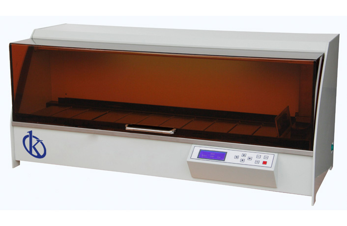
At Kalstein we are MANUFACTURERS, so you can BUY everything you need for your pathology laboratory at excellent PRICES. That is why we invite you to take a look at the HERE