An ultrasound scanner is medical equipment that provides images of most soft tissues without subjecting patients to ionizing radiation. In other words, it allows obtaining diagnostic images from the echoes obtained by the emission of ultrasound waves (the most common). A small instrument called a transducer, very similar to a microphone, emits ultrasound waves. These high-frequency sound waves are transmitted to the area of the body under study and their echo is received. The transducer picks up the echo from the sound waves and a computer converts it into an image that appears on the screen.
Ultrasounds are high frequency sound waves (more than 20,000 cycles per second or 20 kHz) that are not audible to humans. Different tissues alter the waves in different ways, some reflect it directly and others scatter them as echoes before they reach the transducer. Deeper reflected echoes are more attenuated than shallow ones. When the echoes return to the transducer it is possible to reconstruct a two-dimensional map of the tissues.
What are the medical applications of ultrasound scanner?
They are typically used in the radiology department and other clinical departments, as well as in independent imaging centers and private medical practices, primarily for vascular and gynecological-obstetric applications. Some systems include additional transducers to facilitate more specialized diagnostic procedures, such as cardiac, vascular, endovaginal, endorectal, or small part (eg, thyroid, breast, scrotum, prostate) scanning.
How does ultrasound work and what data does it provide?
Ultrasound refers to sound waves emitted at frequencies above the range of human hearing. Frequencies ranging from 2 to 15 megahertz (MHz) are typically used for diagnostic imaging. Ultrasound waves are mechanical (acoustic) vibrations that require a transmission medium and, as they exhibit the normal properties of reflection, refraction, and diffraction of waves, they can be predictably directed, focused, and reflected.
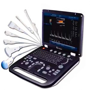
A typical ultrasonic scanning system consists of a beamformer, a central processing unit, a user interface (keyboard, control panel, trackball), several probes (transducers or scan heads), one or more video displays, some kind of recording device and a power supply system.
For ultrasonic imaging, a probe is placed on the skin (after applying an acoustic coupling gel) or inserted into a body cavity. Ultrasonic probes contain one or more elements made of piezoelectric materials (materials that convert electrical energy into acoustic energy and vice versa). When the ultrasonic energy emitted from the probe is reflected by the tissue, the transducer receives some of these reflections (echoes) and converts them back to electrical signals. These signals are processed and transformed into an image (sonogram). Lower sound frequencies provide lower resolution but offer greater tissue penetration, while higher frequencies improve resolution when deep penetration is not required (for example, in pediatric or small-part studies).
At Kalstein we are MANUFACTURERS of high quality medical equipment and at the best PRICES on the market, that is why we present you a new ultrasound scanner and invite you to take a look at HERE