Ultrasound transmits internal images of the body through waves that reflect the mechanical structures of the equipment. It is used to diagnose the causes of any pain in patients, infections, and inflammations, in addition to controlled studies in pregnant patients, to evaluate the brain, hips, and spine.
Also, it reinforces to guide biopsies, determine heart conditions, and assess damage after heart attack. Ultrasound is safe, non-invasive, and does not use radiation. This medium requires little or no preparation.
It develops, by using information-transmitting probes, placed on the skin of patients, and with the help of a computer, creates the image. In addition, ultrasound tests do not use radiation (X-rays), and can show blood flowing through blood vessels.
It should be noted that by studying ultrasound in a conventional way, images are shown in thin and flat fractions of the body and thanks to advances in ultrasound technology, three-dimensional ultrasound (3-D) is included, capable of transforming acoustic wave data into 3-D images. As well as Doppler ultrasounds, it can be part of an ultrasound examination.
Procedures of Ultrasound
The operating mechanisms of ultrasound are developed by acoustic waves that, when measured, cause an echo capable of transforming it into shape, size and consistency. This is added, if it is a solid object or if it contains fluid.
Health professionals use these devices to analyze and visualize the organs, tissues, abnormal masses, tumors, etc. Such images, the shot in the form of a photograph, as well as, can record videos.
On the other hand, Doppler ultrasound is a special ultrasound practice that values the movement of materials within the body. There are three types of Doppler ultrasound:
- The color Doopler, is performed with the help of a computer to transform the Doppler measurements into a set of colors showing the speed and direction of blood flow, through a blood vessel.
- The Doppler with energy provides more details of blood flow, especially in the vessels inside the organs.
- The spectral Doppler presents blood flow controls graphically, based on the path per unit of time and can convert blood flow information, into a distinctive sound that can be heard with each heartbeat.
Interpretation of Ultrasound
The professional in charge of supervising and analyzing ultrasound examinations is known as a radiologist. The provider provides printed images or videos (as the case may be) to the treating physician, or failing that, depends on the medical specialty, where this device is used. In some cases, the specialist may explain the results and use follow-up as part of any necessary treatment or control, in favor of the patient’s health.
It should be noted that specialists must ensure that ultrasound procedures and equipment are safe, comply with the rules, provide high quality images, as well as the associated risks to the patient or staff.
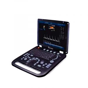
Ultrasound Scanner in Kalstein
We at Kalstein are trained to provide you with all the advanced technology, of our various medical equipment, to enforce all the demands of our users. And in that particular, offer a wide range of ultrasound scanner equipment, which belong to the YR models, they feature 15-inch color LED display with real Doppler function, USB ports and VGA port and 2 probe connectors. Built-in battery, which can be contentiously worked for at least 3 hours when powered off. Primary Application: Abdomen / Cardiac / Obstetrics / Gynecology / Urology / Andrology / Small Parts / Vascular / Pediatrics / Skeletal Muscle and so on .Doppler color based PC with Windows system, which can connect with any printer of any brand. The printed area is adjustable, which can be images, reports or image + report, etc. Built-in DICOM 3.0 protocol.
Visit our website here in this way you can enjoy the best prices and the best advice for your purchase, besides being manufacturers we offer you the best and advanced technology. For more information, check out HERE