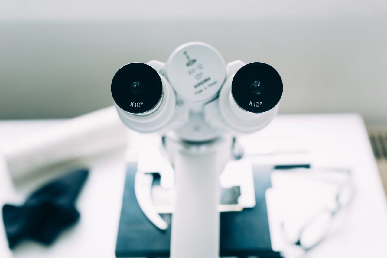
The phase contrast microscope is a type of microscope that allows you to observe live samples without using staining techniques. This microscope is based on the existence of a phase difference between the different light waves that pass through the sample to generate the image of the observation.
How did this microscope originate?
Phase contrast microscopy was invented by the Dutch physicist Frits Zernike in 1932. This great invention earned him the Nobel Prize in Physics in 1953. The main difficulty in observing living cells was that they are practically transparent. For this reason, if they were looked at through a conventional microscope with transmitted light, it was very difficult or even impossible to observe their microscopic details and structures. The usual solution to this problem was to use staining techniques.
The problem in the use of dyes is that their addition is often incompatible with the life of the cells. For this reason, although they are suitable for observing dead cells, their use is often limited when the cells are to be kept alive.
For this type of observations, the phase contrast microscope was created. This microscope manipulates light in such a way that it is possible to increase the contrast of the observed sample. In this way it is possible to observe structures that are invisible through a conventional microscope.
What are the applications of this microscope?
The phase contrast microscope is an instrument to be able to observe all kinds of live samples (cells, microorganisms, tissues) without the need to use dyes. The invention of this microscope resulted in great advances in the field of biology since it allowed the observation of biological processes unknown until now.
Operation of the phase contrast microscope
The phase contrast microscope works in a similar way to a conventional compound microscope but also includes some additional elements that allow it to detect changes in the phases of waves.
First of all there is a spotlight or light source that illuminates the sample. When this light passes through the sample, its waves are affected in different ways. Consequently, these light waves are divided into two parts, known as illumination light and scattered light.
Illumination light is light that passes through the sample without undergoing any change. The rest of the waves, known as scattered light, undergo a phase change due to the refractive index of the specimen in the sample. This is because light travels slightly slower as it passes through the specimen. Consequently, when the light exits the sample there is a phase difference between the waves of the illumination light and those of the scattered light.
This microscope takes advantage of the small differences in refractive indices in different parts of a cell and in different parts of a tissue sample. Light passing through regions of higher refractive index is deflected and out of phase with the main beam of light waves that passed the sample. It pairs other wavelengths out of phase by means of a series of lens and condenser optical rings, cancels out the amplitude of the initial out-of-phase portion of the light beam, and produces useful contrast on the image. The dark parts of the image correspond to the dense portions of the specimen; the light parts of the image correspond to less dense portions.
At Kalstein we provide you with microscopes of the highest technology and quality. That is why we invite you to take a look at the Products menu. HERE